Empirical Force Fields for Biologically Active Divalent Metal Cations in Water
C. Satheesan Babu, and Carmay Lim
J. Phys. Chem. A 2006, 110, 691-699
Informações:
Raio de van der Waals para o Cu2+
Este método não descreve a distorção Jahn Teller, o efeito do elétron desemparelhado do cobre alargando as distâncias axiais ou equatoriais.
vdW parameters for Cu2+
e Rmin/2 deldelG CN Cu-O
(kcal/mol) (angstron) (kcal/mol) # (angstron)
Model A 0.0734 0.8685 -13.3 (-0.1) 6 1.90
Model B 0.1305 1.0763 -13.5 (-0.3) 6 2.03
Model C 0.0427 1.0330 -59.9 (+1.0) 6 1.94
Qual usar? Seria melhor utilizar o modelo que descreve melhor os parâmetros estruturais:
A escolha foi a do modelo C.
Preciso fazer a conversão para o vdW do Cu2+?? Já que este é para o sistema Cu-H2O?????
Observe que no arquivo parmtop uso o Rmin/2 como está listado na tabela acima
Discussion about CuH2O structural geometry
from J. Phys. Chem. A, Vol. 114, No. 35, 2010
Development and Validation of a ReaxFF Reactive Force Field for Cu Cation/Water
Interactions and Copper Metal/Metal Oxide/Metal Hydroxide Condensed Phases
For a long time, it was generally accepted that the copper(II) ion is 6-fold coordinated in aqueous solution with a tetragonally distorted octahedral structure, as would be predicted from the Jahn-Teller effect for a d9 electronic configuration.
This was challenged in 2001 by Pasquarello et al. who proposed a 5-fold coordination based on first-principles MD and neutron diffraction.[10] Their simulation indicated frequent exchanges of square pyramidal and trigonal bipyramidal clusters with five equal Cu-O distances. One year later Persson et al. performed extensive EXAFS and large angle X-ray
scattering (LAXS) experiments and again suggested that the ion was 6-fold coordinated.[14] They modeled the copper-water cluster using several different geometrical configurations and showed that a tetragonally distorted octahedron gave the best fit to the experimental data. In 2003, a quantum-mechanical/molecularmechanical (QM/MM) study by Schwenk et al. also indicated a 6-fold coordination with Jahn-Teller distortion.[12,13] However, another Car-Parrinello molecular dynamics (CPMD) simulation performed by Amira et al.[4] and analysis of the XANES portion of the XAS spectra by Benfatto et al.[15] and Frank et al.[16] suggested that the hydrated copper(II) ion can be better represented as a five-coordinate square pyramidal structure with one elongated axial water molecule.
More recently, Chaboy et al. performed XANES and EXAFS spectroscopy of copper ion hydration [17,18] and argued that “neither the classical Jahn-Teller geometry nor other six- or fivefold coordinated geometries may be proposed unambiguously as the single preferred structure in solution”.[18] According to them, a dynamic view of different geometries exchanging with each other is the right description of copper ion hydration. A review of the structural parameters of Cu2+ aqueous complexes is referenced.[19]
MASS
> CU 63.55
>
> BOND
> nb-CU 75.582 2.027
> o-CU 84.879 1.907
>
> ANGLE
> ha-ca-nb 52.970 116.000 same as ha-ca-n2
> ca-nb-CU 50.523 114.913 from g09 calculations
>
> DIHE
> ca-nb-cp-ca 1 4.800 180.000 2.000
> ca-nb-cp-cp 1 4.800 180.000 2.000
> nb-CU-nb-cp 1 14.800 180.000 2.000
>
> IMPROPER
> o- o-CU-nb 1.1 180.0 2.0
>
> NONBON
> CU 1.0330 0.0427 LJ parms from
> http://dx.doi.org/10.1021/jp054177x
Pesquisar este blog
domingo, 1 de setembro de 2013
Enim - amber
# Check the parameters not included
parmchk -i case.prep -f prepi -o case.frcmod
parmchk -i case.mol2 -f mol2 -o case.frcmod
# Check in mol2 file whether the total charge is the expected one:
cat case_RESP.mol2 | awk '{ sum+=$9} END {print sum}'
1.9996
# Add the remaining charge to be equal to 2.00
# Observe that gaff does not recognize Copper atom
more $AMBERHOME/dat/leap/parm/gaff.dat
# Check the parameters not included in the frcmod file
This command provides a frcmod file:
remark goes here
MASS
Cu 63.6 ATTN, need revision
NONBON
Cu 1.2000 0.0500 ATTN, need revision
Run leap
tleap -f leaprc.ff99SB
source leaprc.gaff
LIG = loadmol2 case.mol2
loadamberparams case.frcmod
saveoff LIG case.lib
saveamberparm LIG case.top case.inpcrd
addions LIG Cl- 0
saveoff LIG case_CI.lib
saveamberparm LIG case_CI.top case_CI.inpcrd
solvatebox LIG TIP3PBOX 14
saveoff LIG case_solv.lib
saveamberparm LIG case_solv.top case_solv.inpcrd
tleap -f leaprc.gaff
source leaprc.ff99SB
loadamberprep case.prep
5) Run sander
mpirun -np 8 sander.MPI -O -i min1.in -o min1.out -p case_solv.top -c case_solv.inpcrd -r case_solv_min1.rst -ref case_solv.inpcrd &
mpirun -np 8 sander.MPI -O -i min2.in -o min2.out -p case_solv.top -c case_solv_min1.rst -r case_solv_min2.rst &
mpirun -np 8 sander.MPI -O -i md1.in -o md1.out -p case_solv.top -c case_solv_min2.rst -r case_solv_md1.rst -x case_solv_md1.mdcrd -ref case_solv_min2.rst &
mpirun -np 8 sander.MPI -O -i md2.in -o md2.out -p case_solv.top -c case_solv_md1.rst -r case_solv_md2.rst -x case_solv_md2.mdcrd &
http://ambermd.org/antechamber/antechamber.html
parmchk -i case.prep -f prepi -o case.frcmod
parmchk -i case.mol2 -f mol2 -o case.frcmod
cat case_RESP.mol2 | awk '{ sum+=$9} END {print sum}'
1.9996
# Add the remaining charge to be equal to 2.00
# Observe that gaff does not recognize Copper atom
more $AMBERHOME/dat/leap/parm/gaff.dat
# Check the parameters not included in the frcmod file
This command provides a frcmod file:
remark goes here
MASS
Cu 63.6 ATTN, need revision
NONBON
Cu 1.2000 0.0500 ATTN, need revision
Run leap
tleap -f leaprc.ff99SB
source leaprc.gaff
LIG = loadmol2 case.mol2
loadamberparams case.frcmod
saveoff LIG case.lib
saveamberparm LIG case.top case.inpcrd
addions LIG Cl- 0
saveoff LIG case_CI.lib
saveamberparm LIG case_CI.top case_CI.inpcrd
solvatebox LIG TIP3PBOX 14
saveoff LIG case_solv.lib
saveamberparm LIG case_solv.top case_solv.inpcrd
tleap -f leaprc.gaff
source leaprc.ff99SB
loadamberprep case.prep
5) Run sander
mpirun -np 8 sander.MPI -O -i min1.in -o min1.out -p case_solv.top -c case_solv.inpcrd -r case_solv_min1.rst -ref case_solv.inpcrd &
mpirun -np 8 sander.MPI -O -i min2.in -o min2.out -p case_solv.top -c case_solv_min1.rst -r case_solv_min2.rst &
mpirun -np 8 sander.MPI -O -i md1.in -o md1.out -p case_solv.top -c case_solv_min2.rst -r case_solv_md1.rst -x case_solv_md1.mdcrd -ref case_solv_min2.rst &
mpirun -np 8 sander.MPI -O -i md2.in -o md2.out -p case_solv.top -c case_solv_md1.rst -r case_solv_md2.rst -x case_solv_md2.mdcrd &
http://ambermd.org/antechamber/antechamber.html
Simulação Cu(H2O)
Teste realizado com dinâmica molecular clássica com os seguintes parâmetros não ligados de XXns
(500 frames gravados)
NONBON
Cu 1.2000 0.05000 ATTN, need revision
Carga 2+
(500 frames gravados)
NONBON
Cu 1.2000 0.05000 ATTN, need revision
Carga 2+
![]() |
| g(r) Cobre- Cloreto |
![]() |
| g(r) Cobre - Oagua |
Cu_Cl_2.dat Media (Desvio): 2.11 0.08
Cu_Cl_3.dat Media (Desvio): 2.10 0.08
Cu_solv189.dat Media (Desvio): 2.16 0.14
Cu_solv297.dat Media (Desvio): 2.15 0.14
![]() |
| g(r) Copper+Ow |
![]() |
| g(r) Copper+Cl- |
Simulação DNA+Cuisaepy+
# Copiar modelo que será utilizado no docking
tleap -f leaprc.gaff
source leaprc.ff99SB
# CARREGAR O COMPLEXO DE COBRE (o loadoff deve vir antes do loadpdb !!!!!!)
loadoff cie.lib
CIE = loadpdb cie.pdb
loadamberparams cie.frcmod
check CIE
# CARREGAR O DNA
DNA = loadmol2 3GC.mol2
savepdb DNA DNA.pdb
check DNA
# COMBINAR OS DOIS RESIDUOS
CIED = combine { CIE DNA }
# COLOCAR ci, SOLVATACAO E SALVAR
saveamberparm CIED cie.top cie.crd
savepdb CIED cied.pdb
addions CIED Na+ 0
saveamberparm CIED cie_ci.top cie_ci.crd
solvatebox CIED TIP3PBOX 14
saveamberparm CIED cie_solv.top cie_solv.crd
savepdb CIED cie_solv.pdb
tleap -f leaprc.gaff
source leaprc.ff99SB
# CARREGAR O COMPLEXO DE COBRE (o loadoff deve vir antes do loadpdb !!!!!!)
loadoff cie.lib
CIE = loadpdb cie.pdb
loadamberparams cie.frcmod
check CIE
# CARREGAR O DNA
DNA = loadmol2 3GC.mol2
savepdb DNA DNA.pdb
check DNA
# COMBINAR OS DOIS RESIDUOS
CIED = combine { CIE DNA }
# COLOCAR ci, SOLVATACAO E SALVAR
saveamberparm CIED cie.top cie.crd
savepdb CIED cied.pdb
addions CIED Na+ 0
saveamberparm CIED cie_ci.top cie_ci.crd
solvatebox CIED TIP3PBOX 14
saveamberparm CIED cie_solv.top cie_solv.crd
savepdb CIED cie_solv.pdb
__________________
Foram realizadas três dinâmicas: model1, model4 e model5 (fazer). Os testes foram realizados no i7 e no cenapad
Modelo 4 (i7)
Em todos os testes o complexo se desligou do DNA. Motivos:
1) Erro na parametrização da carga do complexo(?)
2) DNA muito pequeno?
_____________________________
OBS:
Para rodar com o cuda, use primeiro o MPI na minimização e faça um pouco da dinâmica (md2.in) com MPI também. Assim quand tiver uma oa estrutura inicie o cálculo com gpu.
Observe que o complexo pode ficar emparelhado com o backbone com a região da piridina ou a região do oxindol. Observe que o =O o oxindol é bastante "pontiagudo" o que parece dificultar a interação com o DNA
Estudo 6DNA (12 bases) com a sequência AT ligado ao [Cu(isaepy(H2O)]+
Trajetória ~ ns
BOX: 24
(Cenapad - CUDA)
O sistema DNA apresentou bastante vibração. Ocorreu a separação da última sequência AT. A Timina se ligou om a adenina através do =O que não faz HB com o NH2 da adenina. Ocorreu também a interação entre bases de planos distintos.
O complexo foi "docado" com o cobre para o lado do solvente. O complexo se manteve estável a menos de algumas vibrações com a tentativa de trocar de sequência. Não houve a possibilidade de sair do DNA.
Estudo 3DNA (6 bases) com a sequência GC ligad ao [Cu(isaenim)]+ (forma enol).
Trajetória de ~56 ns
(Cenapad - CUDA)
BOX: 27
O composto inicialmente foi 'docado' com o cobre voltado para o DNA. O composto se desliga ficando preso pelo amino do imidazol. Em seguida o composto se vira e se liga com o lado hidrofóbico.
O DNA se mantém bem estruturado, sem grandes variações estruturais.
Estudo 6DNA (12 bases) com a sequência CG ligado ao [Cu(isaepy(H2O)]+
Trajetória ~ 54 ns
BOX: 24
Foram realizadas três dinâmicas: model1, model4 e model5 (fazer). Os testes foram realizados no i7 e no cenapad
Modelo 4 (i7)
Em todos os testes o complexo se desligou do DNA. Motivos:
1) Erro na parametrização da carga do complexo(?)
2) DNA muito pequeno?
_____________________________
OBS:
Para rodar com o cuda, use primeiro o MPI na minimização e faça um pouco da dinâmica (md2.in) com MPI também. Assim quand tiver uma oa estrutura inicie o cálculo com gpu.
Observe que o complexo pode ficar emparelhado com o backbone com a região da piridina ou a região do oxindol. Observe que o =O o oxindol é bastante "pontiagudo" o que parece dificultar a interação com o DNA
Estudo 6DNA (12 bases) com a sequência AT ligado ao [Cu(isaepy(H2O)]+
Trajetória ~ ns
BOX: 24
(Cenapad - CUDA)
O sistema DNA apresentou bastante vibração. Ocorreu a separação da última sequência AT. A Timina se ligou om a adenina através do =O que não faz HB com o NH2 da adenina. Ocorreu também a interação entre bases de planos distintos.
O complexo foi "docado" com o cobre para o lado do solvente. O complexo se manteve estável a menos de algumas vibrações com a tentativa de trocar de sequência. Não houve a possibilidade de sair do DNA.
Estudo 3DNA (6 bases) com a sequência GC ligad ao [Cu(isaenim)]+ (forma enol).
Trajetória de ~56 ns
(Cenapad - CUDA)
BOX: 27
O composto inicialmente foi 'docado' com o cobre voltado para o DNA. O composto se desliga ficando preso pelo amino do imidazol. Em seguida o composto se vira e se liga com o lado hidrofóbico.
O DNA se mantém bem estruturado, sem grandes variações estruturais.
Estudo 6DNA (12 bases) com a sequência CG ligado ao [Cu(isaepy(H2O)]+
Trajetória ~ 54 ns
BOX: 24
(Cenapad: CUDA)
O complexo fica muito tempo preso ao backbone através da 5 posição de coordenação do cobre. Neste composto deixei uma água sempre fixa ao complexo. A última base também se desligou temporariamente.
Estudo 3DNA (6 bases) com a sequência CG ligado ao [Cu(isaepy(H2O)]+
Trajetória ~ ns
BOX: 24
Trajetória ~ ns
BOX: 24
(Cenapad: CUDA)
O composto fica bem mais instável, se desligando e religando ao DNA diversas vezes
O composto tenta se inserir entre as bases (intercalação) através da piridina. Também ocorre a interação frequente com o backbone através da 5 posição do cobre
A interação é mais frequente com o lado hidrofóbico, lembrando que neste modelo há uma água fixa ao composto.
[Cu(isaepy)]+ cotovelos
Comparação entre os resultados do docking para diferentes 'cotovelos'
Simulação feita com o Autodock vina e este atribui o seu campo eletrostático sem considerar as cargas fornecidas pelo usuário.
Observe que ainda não fiz tais testes com a dinâmica. E as que fiz foi com a forma 1

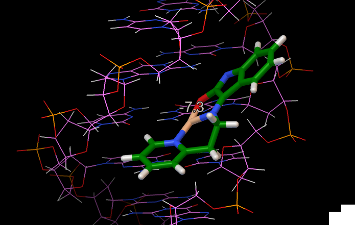
Resultados Docking Forma 2:
Em resumo
a Forma 1 pode intercalar pelo oxindol do lado do cobre ou pela piridina do lado invertido
a Forma 2 pode intercalar pela piridina pelo lado do cobre ou pelo oxindol do lado invertido.
Utilizei as Gasteiger partial atomic charges e como não há cargas para o cobre adicionei carga +1. Abaixo segue a atribuição das cargas para os sítios mais reativos. Entretanto como pode ser lido abaixo, os metais são tratados como doadores de ligação de hidrogênio e estas cargas são ignoradas no cálculo vina.
Cargas obtidas através do autodock
1)
ATOM 3 CU LIG d 1 4.600 8.650 2.925 0.00 0.00 1.000 Cu
ATOM 1 N LIG d 1 6.507 8.273 2.448 0.00 0.00 -0.266 N
ATOM 2 N LIG d 1 4.527 7.352 4.429 0.00 0.00 -0.284 N
ATOM 19 N LIG d 1 1.429 8.700 5.283 0.00 0.00 -0.220 NA
ATOM 20 O LIG d 1 2.874 9.254 3.523 0.00 0.00 -0.168 OA
2)
ATOM 1 CU LIG d 1 4.487 8.832 3.005 0.00 0.00 1.000 Cu
ATOM 2 N LIG d 1 5.870 7.908 1.890 0.00 0.00 -0.266 N
ATOM 4 N LIG d 1 4.425 7.427 4.411 0.00 0.00 -0.284 N
ATOM 3 N LIG d 1 1.614 9.025 5.712 0.00 0.00 -0.220 NA
ATOM 5 O LIG d 1 2.954 9.599 3.877 0.00 0.00 -0.168 OA
Simulação feita com o Autodock vina e este atribui o seu campo eletrostático sem considerar as cargas fornecidas pelo usuário.
Observe que ainda não fiz tais testes com a dinâmica. E as que fiz foi com a forma 1
Resultados do docking da Forma 1:

Resultados Docking Forma 2:
Em resumo
a Forma 1 pode intercalar pelo oxindol do lado do cobre ou pela piridina do lado invertido
a Forma 2 pode intercalar pela piridina pelo lado do cobre ou pelo oxindol do lado invertido.
Utilizei as Gasteiger partial atomic charges e como não há cargas para o cobre adicionei carga +1. Abaixo segue a atribuição das cargas para os sítios mais reativos. Entretanto como pode ser lido abaixo, os metais são tratados como doadores de ligação de hidrogênio e estas cargas são ignoradas no cálculo vina.
Cargas obtidas através do autodock
1)
ATOM 3 CU LIG d 1 4.600 8.650 2.925 0.00 0.00 1.000 Cu
ATOM 1 N LIG d 1 6.507 8.273 2.448 0.00 0.00 -0.266 N
ATOM 2 N LIG d 1 4.527 7.352 4.429 0.00 0.00 -0.284 N
ATOM 19 N LIG d 1 1.429 8.700 5.283 0.00 0.00 -0.220 NA
ATOM 20 O LIG d 1 2.874 9.254 3.523 0.00 0.00 -0.168 OA
2)
ATOM 1 CU LIG d 1 4.487 8.832 3.005 0.00 0.00 1.000 Cu
ATOM 2 N LIG d 1 5.870 7.908 1.890 0.00 0.00 -0.266 N
ATOM 4 N LIG d 1 4.425 7.427 4.411 0.00 0.00 -0.284 N
ATOM 3 N LIG d 1 1.614 9.025 5.712 0.00 0.00 -0.220 NA
ATOM 5 O LIG d 1 2.954 9.599 3.877 0.00 0.00 -0.168 OA
"AutoDock Vina ignores the user-supplied partial charges. It has its own way of dealing with the electrostatic interactions through the hydrophobic and the hydrogen bonding terms."
"Following Xscore, we formally treat metals as hydrogen bond donors"
quinta-feira, 1 de agosto de 2013
Glycans - complexo de açúcares
Three major repeating biomacromolecules,
O ser humano possui aprox. 25.000 genes enquanto uma bactéria unicelular possui aprox. 6.000 genes. Apensar da aparente pequena quantidade de genes humanos comparados a uma bactéria, a diferença está na "pós-translacional modificação" das proteínas no ser humano. Uma das mais comuns modificações está na glicolização de proteínas
O estudo dos açúcares que influenciam as funções das proteínas e lipídios é chamado de glicoma. Em uma célula sadia o glicoma tem certas características diferentes de uma célula cancerígena.
Os glicans são formados no Retículo endoplamático (ER) e/ou(?) complexo de Golgi e assim os glicans se ligam as proteínas.
Diferente do DNA que tem uma sequência única de formação 3'-5' e as proteínas que tem o N-terminal e o XX-inicial, os sacarídeos podem se formar de maneiras diversas dependendo de suas conecções.
Imagens do vídeo:
http://www.youtube.com/watch?v=WCbg-kOY_8E
http://ibioseminars.hhmi.org/lectures/chemicalbiologybiophysics/carolyn-bertozzi.html
N-Acetylgalactosamine (GalNAc), is an amino sugar derivative of galactose. (componente do O-glycan)
N-Acetylglucosamine (N-acetyl-D-glucosamine, or GlcNAc, or NAG) is a monosaccharide derivative of glucose. (Componente do N-glycan)
Os carboidratos das proteínas da superfície celular tem duas funções dependendo de sua estrutura:
- polynucleotides,
- polypeptides and
- carbohydrates,
Encoding and transmission of information relies on the construction of diverse macromolecules that contain the message.
Polynucleotides serve as the blueprint of life in form of DNA;
polypeptides carry out most of reactions in living cells.
Both polymers are strictly linear and derived biosynthetically via reliable templated syntheses.
DNA is composed of 4 nucleotides and mammalian proteins have 20 proteinogenic amino acids that determine polymer diversity: 4096 (46) hexanucleotides and 64 million (206) hexapeptides are possible.
Posttranslational modifications such as
- phosphorylation,
- glycosylation, and
- lipidation
O ser humano possui aprox. 25.000 genes enquanto uma bactéria unicelular possui aprox. 6.000 genes. Apensar da aparente pequena quantidade de genes humanos comparados a uma bactéria, a diferença está na "pós-translacional modificação" das proteínas no ser humano. Uma das mais comuns modificações está na glicolização de proteínas
O estudo dos açúcares que influenciam as funções das proteínas e lipídios é chamado de glicoma. Em uma célula sadia o glicoma tem certas características diferentes de uma célula cancerígena.
Os glicans são formados no Retículo endoplamático (ER) e/ou(?) complexo de Golgi e assim os glicans se ligam as proteínas.
Diferente do DNA que tem uma sequência única de formação 3'-5' e as proteínas que tem o N-terminal e o XX-inicial, os sacarídeos podem se formar de maneiras diversas dependendo de suas conecções.
Mucin | ||
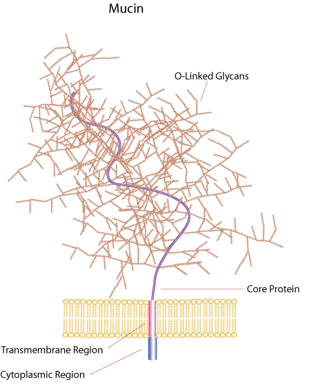
Mucins are a diverse family of densely glycosylated proteins. Mucin domains within the protein core are rich in threonine, serine and hydroxyproline enabling post-translational O-glycosylation. The highly glycosylated properties of mucins make them resistant to proteolysis and able to hold water, giving them the gel-like properties found in mucosal barriers. Mucins also contain cysteine-rich regions that participate in intermolecular cross-linking and are typically secreted as large aggregates. Mucins may also be associated with membranes and may serve as receptor-like ligands for carbohydrate-binding molecules.
![]() |
| http://www.sigmaaldrich.com/life-science/metabolomics/enzyme-explorer/learning-center/structural-proteins/mucin.html |
Imagens do vídeo:
Carolyn Bertozzi (UC Berkeley) Part 1: Chemical Glycobiology
ibiology
http://www.youtube.com/watch?v=WCbg-kOY_8E
http://ibioseminars.hhmi.org/lectures/chemicalbiologybiophysics/carolyn-bertozzi.html
![]() |
| Glico proteínas e glico lipídios |
![]() |
| 9 monossacarídios encontrado nos vertebrados |
![]() |
| Sacarídeos nas células sanguíneas que diferenciam os tipos O, A, B e AB. Assim como as enzimas que as formam são diferentes. |
![]() |
| N-glycan core |
N-Acetylgalactosamine (GalNAc), is an amino sugar derivative of galactose. (componente do O-glycan)
![]() |
| GalNAc |
N-Acetylglucosamine (N-acetyl-D-glucosamine, or GlcNAc, or NAG) is a monosaccharide derivative of glucose. (Componente do N-glycan)
![]() |
| GlcNAc |
Os carboidratos das proteínas da superfície celular tem duas funções dependendo de sua estrutura:
- Promover a metástase
- Inibir a metástase
Estes são classificados por sua ligação com a proteína:
N-glycans - ligados a asparagina (ASN) N
O-glycans - ligados a serina (SER) S ou a Threonine (THR) T
Os O-glycans em mamíferos são classificados em 4 grupos (core1 - core4)
Papers DNA+Cu-complexes
1)
Metal complex–DNA binding: Insights from molecular dynamics and DFT/MM calculations
http://www.sciencedirect.com/science/article/pii/S0162013413000688
2)
Molecular recognition between DNA and a copper-based anticancer complex
http://pubs.rsc.org/en/Content/ArticleLanding/2012/CP/c2cp42185b#!divAbstract
3)
Synthesis and crystal structure of a ternary copper(II) complex of 2,2’-bipyridine and picrate: Molecular docking, reactivity towards DNA and in vitro anticancer activity
http://www.sciencedirect.com/science/article/pii/S0022286013009289
Syntheses and crystal structures of tetracopper(II) complexes bridged by asymmetric N,N′-bis(substituted)oxamides: Molecular docking, DNA-binding and in vitro anticancer activity
http://www.sciencedirect.com/science/article/pii/S0162013413001918
Intercalative interaction of asymmetric copper(II) complex with DNA: Experimental, molecular docking, molecular dynamics and TDDFT studies
http://www.sciencedirect.com/science/article/pii/S0162013413001980
Theoretical Simulations on Interactions of Mono- and Dinuclear Metallonucleases with DNA
http://pubs.acs.org/doi/abs/10.1021/jp306998f?journalCode=jpcbfk
4)
Evaluation of Amber force field parameters for copper(II) with pyridylmethyl-amine and benzimidazolylmethyl-amine ligands: A quantum chemical study
http://www.sciencedirect.com/science/article/pii/S0009261408003382
5)
Copper−1,10-Phenanthroline Complexes Binding to DNA: Structural Predictions from Molecular Simulations
http://pubs.acs.org/doi/abs/10.1021/jp901210g
ver
http://pubs.rsc.org/en/content/articlelanding/2010/ic/b918406f#!divAbstract
http://pubs.acs.org/doi/abs/10.1021/cr400135x
Metal complex–DNA binding: Insights from molecular dynamics and DFT/MM calculations
http://www.sciencedirect.com/science/article/pii/S0162013413000688
2)
Molecular recognition between DNA and a copper-based anticancer complex
http://pubs.rsc.org/en/Content/ArticleLanding/2012/CP/c2cp42185b#!divAbstract
3)
Synthesis and crystal structure of a ternary copper(II) complex of 2,2’-bipyridine and picrate: Molecular docking, reactivity towards DNA and in vitro anticancer activity
http://www.sciencedirect.com/science/article/pii/S0022286013009289
Syntheses and crystal structures of tetracopper(II) complexes bridged by asymmetric N,N′-bis(substituted)oxamides: Molecular docking, DNA-binding and in vitro anticancer activity
http://www.sciencedirect.com/science/article/pii/S0162013413001918
Intercalative interaction of asymmetric copper(II) complex with DNA: Experimental, molecular docking, molecular dynamics and TDDFT studies
http://www.sciencedirect.com/science/article/pii/S0162013413001980
Theoretical Simulations on Interactions of Mono- and Dinuclear Metallonucleases with DNA
http://pubs.acs.org/doi/abs/10.1021/jp306998f?journalCode=jpcbfk
4)
Evaluation of Amber force field parameters for copper(II) with pyridylmethyl-amine and benzimidazolylmethyl-amine ligands: A quantum chemical study
http://www.sciencedirect.com/science/article/pii/S0009261408003382
5)
Copper−1,10-Phenanthroline Complexes Binding to DNA: Structural Predictions from Molecular Simulations
http://pubs.acs.org/doi/abs/10.1021/jp901210g
ver
http://pubs.rsc.org/en/content/articlelanding/2010/ic/b918406f#!divAbstract
http://pubs.acs.org/doi/abs/10.1021/cr400135x
Ligantes orgânicos de DNA
Quais são as perguntas e dúvidas sobre a interação de drogas (intercalantes ou não em DNA?)
Resistance to chemotherapy and molecularly targeted therapies is a major problem facing current cancer research
In recent years, the general consensus on the crucial role of the interactions of metallo drugs with proteins indetermining the compounds’ pharmacological action, uptake and biodistribution, as well
as their overall toxicity profile, resulted in an exponential increase in the number of studies
Quais são os principais grupos que procuram responder a estas perguntas?
Quais são as técnicas experimentais e computacionais pra auxiliar neste problema?
Incluir uma tabela com o deltaG dos compostos
Porque os intergalantes tem preferência por sequências CG?
__________________________________________________________
Ethidium-bromide
Resistance to chemotherapy and molecularly targeted therapies is a major problem facing current cancer research
In recent years, the general consensus on the crucial role of the interactions of metallo drugs with proteins indetermining the compounds’ pharmacological action, uptake and biodistribution, as well
as their overall toxicity profile, resulted in an exponential increase in the number of studies
Quais são os principais grupos que procuram responder a estas perguntas?
Quais são as técnicas experimentais e computacionais pra auxiliar neste problema?
Incluir uma tabela com o deltaG dos compostos
Porque os intergalantes tem preferência por sequências CG?
__________________________________________________________
Ethidium-bromide
![]() |
| Ethidium-bromide |
hoechst_33258
![]() |
| hoechst_33258 |
![]() |
| 1DNH |
![]() |
| 1QSX |
![]() |
| 8BNA |
1DNH (5'-D(*CP*GP*CP*GP*AP*AP*TP*TP*CP*GP*CP*G)-3')
1QSX 5'-D(CP*TP*TP*TP*TP*GP*CP*AP*AP*AP*AP*G)-3'
8BNA (5'-D(*CP*GP*CP*GP*AP*AP*TP*TP*CP*GP*CP*G)-3')
Em laranja estão as regiões onde o hoechst interage com o DNA
Teste com dinâmica molecular: Tomaremos primeiramente a estrutura de NMR 1QSX que possui informação dos hidrogênios. A sequência que estudarei será AAAA.
Qual a carga do ligante? Considerei a carga +1 segundo o paper "Protonation Equilibria of Hoechst 33258 in aqueous solution (Helvetica Chimica Acta vol 88 (2005) pp53
![]() |
| Hoechst da estrutura 1QSX |
![]() |
| O Hoechst interage com o oxigênio da timina e o N da adenina através do grupo amina secundária (estrutura 1QSX) |
Daunomicyn - intercalador - intercala pelo minor groove
1AL9
1D11
308D
482D

________________________________________________________
Preparação do sistema:
antechamber -i hoechst.pdb -fi pdb -o hoechst.mol2 -fo mol2 -c bcc -s 2 -nc 1
parmchk -i hoechst.mol2 -f mol2 -o hoechst.frcmod
# Copiar modelo que será utilizado no docking
#tleap -f leaprc.gaff
source leaprc.ff99SB
# CARREGAR O COMPLEXO DE COBRE (o loadoff deve vir antes do loadpdb !!!!!!)
#loadoff cie.lib
HT = loadmol2 hoechst.mol2
loadamberparams hoechst.frcmod
check HT
# CARREGAR O DNA
DNA = loadpdb DNA.pdb
savepdb DNA DNA.pdb
check DNA
# COMBINAR OS DOIS RESIDUOS
CIED = combine { HT DNA }
# COLOCAR ci, SOLVATACAO E SALVAR
saveamberparm CIED cie.top cie.crd
savepdb CIED cied.pdb
addions CIED Na+ 0
saveamberparm CIED cie_ci.top cie_ci.crd
solvatebox CIED TIP3PBOX 24
saveamberparm CIED cie_solv.top cie_solv.crd
savepdb CIED cie_solv.pdb
Papers
Designer DNA-binding drugs: the crystal structure of a meta-hydroxy analogue of Hoechst 33258 bound to d(CGCGAATTCGCG)2
George R. Clark1,2, Christopher J. Squire2, Emily J. Gray1, Werner Leupin3,+ and
Stephen Neidle
http://nar.oxfordjournals.org/content/24/24/4882.full.pdf+html
Nucleic Acids Research, 1996, Vol. 24, No. 24
Texto que cita a maior importância das interações de van der Waals e contatos hidrofóbicos do que ligações de hidrogênio nos minor groove binders.
Sequence-dependent variation in DNA minor groove width dictates orientational preference of Hoechst 33258 in A-tract recognition: solution NMR structure of the 2:1 complex with d(CTTTTGCAAAAG)
Nucleic Acids Res. 2000 Feb 1;28(3):728-35.
Gavathiotis E, Sharman GJ, Searle MS.
The majority of small molecules that bind in the minor groove of duplex DNA, including distamycin and Hoechst 33258, have a marked preference for AT rich sequences.
Título: Theoretical study of molecular recognition by Hoechst 33258 derivatives
Autor(es): Kakkar, R; Suruchi; Grover, R
Fonte: JOURNAL OF BIOMOLECULAR STRUCTURE & DYNAMICS Volume: 23 Edição: 1 Páginas: 37-47 Publicado: AUG 2005
Número de citações: 6 (de todas as bases de dados)
The factors responsible for the binding of Hoechst 33258 with DNA residues have been investigated in this work using the AMI method. First and foremost, it is found that, although all crystal structure determinations indicate a preference for binding at AT rich sites, the hydrogen bond strength is actually greater for complexes with cytosine and guanine. From this, it has been inferred that other factors such as electrostatic, van der Waals interactions and nonbonded contacts with the walls of the minor groove have a strong role to play in the binding process. The hydrogen bond is found to be stronger for complexation with the thymine O-2 than with the adenine N-3, in line with experimental observations. Combined QM/MM studies on the drug complexed with the Dickerson-Drew dodecamer reveal that binding induces structural changes in both the ligand as well as DNA. Electron donating substituents at the para position in the phenyl ring of Hoechst 3:3258 lead to stronger binding with DNA. A correlation with the octanol/water partition coefficients points to the importance of hydrophobic and electrostatic interactions.
Título: A molecular thermodynamic view of DNA-drug interactions: a case study of 25 minor-groove binders
Autor(es): Shaikh, SA; Ahmed, SR; Jayaram, B
Fonte: ARCHIVES OF BIOCHEMISTRY AND BIOPHYSICS Volume: 429 Edição: 1 Páginas: 81-99 DOI: 10.1016/j.abb.2004.05.019 Publicado: SEP 1 2004
Número de citações: 52 (de todas as bases de dados)
Developing a molecular view of the thermodynamics of DNA recognition is essential to the design of ligands for regulating gene expression. In a first comprehensive attempt at sketching an atlas of DNA–drug energetics, we present here a detailed thermodynamic view of minor-groove recognition by small molecules via a computational study on 25 DNA–drug complexes. The studies are configured in the MMGBSA (Molecular Mechanics-Generalized Born-Solvent Accessibility) framework at the current state of the art and facilitate a structure–energy component correlation. Analyses were conducted on both energy minimized structures of DNA–drug complexes and molecular dynamics trajectories developed for the purpose of this study. While highlighting the favorable role of packing, shape complementarity, and van der Waals and hydrophobic interactions of the drugs in the minor groove in conformity with experiment, the studies reveal an interesting annihilation of favorable electrostatics by desolvation. Structural modifications attempted on the ligands point to the requisite physico-chemical factors for obtaining improved binding energies. Hydrogen bonds predicted to be important for specificity based on structural considerations do not always turn out to be significant to binding in post facto analyses of molecular dynamics trajectories, which treat thermal averaging, solvent, and counterion effects rigorously. The strength of the hydrogen bonds retained between the DNA and drug during the molecular dynamics simulations is ∼1 kcal/mol. Overall, the study reveals the compensatory nature of the diverse binding free energy components, possible threshold limits for some of these properties, and the availability of a computationally viable free energy methodology which could be of value in drug-design endeavors.
Título: DNA-binding of drugs used in medicinal therapies
Autor(es): Bischoff, G; Hoffmann, S
Fonte: CURRENT MEDICINAL CHEMISTRY Volume: 9 Edição: 3 Páginas: 321-348 Publicado: FEB 2002
Número de citações: 42 (de todas as bases de dados)
The interactions of various low-molecular weight substances with DNA are naturally relevant mechanisms in the cellular cycle and so also used in medicinal treatment. Depending on the particular drug structure, DNA-binding modes like groove-binding, intercalating and/or stacking, give rise to supramolecular assemblies of the polynucleotides, as well as influence the DNA-protein binding.
In this review, we compare the underlying molecular structures, including general aspects of DNA sequences, with the benefit in medicinal treatment. While so far interest in this field had mainly been devoted to isolated nucleic acid/drug interactions, the present paper will focus on drug efficiencies generating and influencing supramolecular organizations and their complex sequence-dependent structure-activity codes. In particular, the attention will be directed to stereoelectronic relationships. Spatial enantioselective properties are discussed in details. As examples, the drug self-assemblies, as well as the influence of drugs on supramolecular DNA formations are described. A hypothetical connection between drug-influenced DNA-toroids and the formation of micronuclei in tissues will be interpreted.
Título: Recent developments in sequence selective minor groove DNA effectors
Autor(es): Reddy, BSP; Sharma, SK; Lown, JW
Fonte: CURRENT MEDICINAL CHEMISTRY Volume: 8 Edição: 5 Páginas: 475-508 Publicado: APR 2001
Número de citações: 86 (de todas as bases de dados)
Título: A new class of symmetric bisbenzimidazole-based DNA minor groove-binding agents showing antitumor activity
Autor(es): Mann, J; Baron, A; Opoku-Boahen, Y; et al.
Fonte: JOURNAL OF MEDICINAL CHEMISTRY Volume: 44 Edição: 2 Páginas: 138-144 DOI: 10.1021/jm000297b Publicado: JAN 18 2001
Número de citações: 130 (de todas as bases de dados)
Título: DNA minor-groove recognition by small molecules
Autor(es): Neidle, S
Fonte: NATURAL PRODUCT REPORTS Volume: 18 Edição: 3 Páginas: 291-309 DOI: 10.1039/a705982e Publicado: 2001
Número de citações: 224 (de todas as bases de dados)
Título: Structural studies of atom-specific anticancer drugs acting on DNA
Autor(es): Yang, XL; Wang, AHJ
Fonte: PHARMACOLOGY & THERAPEUTICS Volume: 83 Edição: 3 Páginas: 181-215 DOI: 10.1016/S0163-7258(99)00020-0 Publicado: SEP 1999
Número de citações: 149 (de todas as bases de dados)
Rational recognition of nucleic acid sequences
Hirokazu Iida, Guofeng Jia, J William Lown E-mail the corresponding author
Current Opinion in Biotechnology
Volume 10, Issue 1, 1 February 1999, Pages 29–33
Factors contributing to the molecular recognition of DNA binders
Although the structures of Hoechst 33258 and netropsin are ostensibly quite dissimilar, examination of the molecular recognition surface components of the two ligands directed towards the DNA minor-groove receptor reveals significant similarities.
Based on not only X-ray analysis but also NMR and biological techniques of assessing DNA–minor-groove-binder interactions, progress has been made in understanding some of the factors contributing to the molecular recognition processes:
Antihelicase Action of DNA-Binding Anticancer Agents: Relationship to Guanosine-Cytidine Intercalator Binding
An additional interesting correlation is the antihelicase action of DNA-intercalating antibiotics and their DNA-binding preference for G-C base pair sites.
The G-C base pair binding preference of the intercalating antibiotics may result from evolutionary selection because of the higher G-C binding stability, compared with A-T
binding stability. The combination of the higher base pair stability at G-C regions and increased duplex DNA stability induced by intercalating antibiotic yields a total additive stability of the intercalator-G-C base pair complex that resists helicase action.
DNA-intercalating antibiotics stabilize duplex DNA and increase
the energy required to separate paired DNA strands (i.e.,
DNA melting) (17). The DNA-intercalating antibiotics daunorubicin
(10), actinomycin D (11), elsamicin (12), and echinomycin
(13) bind preferentially at G-C base pairs of duplex
DNA, as do most natural intercalating antibiotics. G-C base
pairs in duplex DNA have much greater electronic complementarity
than do the A-T base pairs and are the most stable of
the hydrogen-bonded base pairs, as seen in DNA melting. A
reasonable assumption is that the maximal DNA helicase catalytic
power is used to separate G-C base pairs. The combination
ofthe high G-C base pair stability and the increased duplex
DNA stability produced by an intercalated antibiotic at a G-C
site increases the overall duplex DNA stability at the intercalator-
G-C locus. This supernormal duplex DNA stability at the
intercalator-G-C locus may exceed the catalytic power exerted
by helicases to affect duplex DNA strand separation and may
thus slow or stop helicase action. The factor of G-C base pair
stability may have helped to determine the evolutionary selection
of intercalating antibiotics for G-C-binding specificity.
Título:CORRELATION OF THE BASE SPECIFICITY OF DNA - INTERCALATING
LIGANDS WITH THEIR PHYSICO-CHEMICAL PROPERTIES
From analysis of eqs. l-6 a number of conclusions
may be drawn concerning the affinity and specificity
of heterocyclic ligands for DNA. The relative hydro-
phobicities of the ligands correlate neither with the
affinity for DNA nor with the base specificity (eqs. 2
and 5). At physiological pH the ligands are present
almost exclusively in the ionised form in which form
they interact with DNA. One would therefore expect
that electronic parameters would be more important
in determining binding. Eq. 1 shows some correlation
between affinity for DNA and charge transfer inter-
action. This correlation is improved slightly by adding
the hydrophobicity parameter (eq. 3). This suggests
that binding affinity for DNA depends on-a number
of different parameters, electronic, hydrophobic and
also possibly steric. Base specificity on the other hand
appears to depend almost exclusively on electronic factors. This is shown by the very good linear correla-
tion between Miiller’s (Y value and the relative charge
transfer affinity constant KGMP (eq. 4) which is not
significantly altered by the addition of the hydro-
phobicity parameter (eq. 6).
Thus we have shown that base specificity in these
ligands is related to their electronic characteristics.
These are in turn related to chemical structure and
IV-methylation increases both base specificity and
relative charge transfer affinity. The precise effect of
slight variations of chemical structure on the electronic 1
characteristics of the molecule and also the importance
of steric parameters in binding affinity and base
specificity needs to be further investigated.
Título:
Cancer drug resistance: an evolving paradigm Resistance to chemotherapy and molecularly targeted therapies is a major problem facing current cancer research
DOI: 10.1093/NAR/28.3.728 - 1QSX
Gavathiotis E, Sharman GJ, Searle MS.
The majority of small molecules that bind in the minor groove of duplex DNA, including distamycin and Hoechst 33258, have a marked preference for AT rich sequences.
Recently it has been suggested that sequence specificity may be dominated by ability to select binding sites on the basis of groove width, allowing optimum van der Waal’s complementarity and hydrophobic surface burial, rather than specific hydrogen bonding interactions with the groove floor
Título: Theoretical study of molecular recognition by Hoechst 33258 derivatives
Autor(es): Kakkar, R; Suruchi; Grover, R
Fonte: JOURNAL OF BIOMOLECULAR STRUCTURE & DYNAMICS Volume: 23 Edição: 1 Páginas: 37-47 Publicado: AUG 2005
Número de citações: 6 (de todas as bases de dados)
The factors responsible for the binding of Hoechst 33258 with DNA residues have been investigated in this work using the AMI method. First and foremost, it is found that, although all crystal structure determinations indicate a preference for binding at AT rich sites, the hydrogen bond strength is actually greater for complexes with cytosine and guanine. From this, it has been inferred that other factors such as electrostatic, van der Waals interactions and nonbonded contacts with the walls of the minor groove have a strong role to play in the binding process. The hydrogen bond is found to be stronger for complexation with the thymine O-2 than with the adenine N-3, in line with experimental observations. Combined QM/MM studies on the drug complexed with the Dickerson-Drew dodecamer reveal that binding induces structural changes in both the ligand as well as DNA. Electron donating substituents at the para position in the phenyl ring of Hoechst 3:3258 lead to stronger binding with DNA. A correlation with the octanol/water partition coefficients points to the importance of hydrophobic and electrostatic interactions.
Título: A molecular thermodynamic view of DNA-drug interactions: a case study of 25 minor-groove binders
Autor(es): Shaikh, SA; Ahmed, SR; Jayaram, B
Fonte: ARCHIVES OF BIOCHEMISTRY AND BIOPHYSICS Volume: 429 Edição: 1 Páginas: 81-99 DOI: 10.1016/j.abb.2004.05.019 Publicado: SEP 1 2004
Número de citações: 52 (de todas as bases de dados)
Developing a molecular view of the thermodynamics of DNA recognition is essential to the design of ligands for regulating gene expression. In a first comprehensive attempt at sketching an atlas of DNA–drug energetics, we present here a detailed thermodynamic view of minor-groove recognition by small molecules via a computational study on 25 DNA–drug complexes. The studies are configured in the MMGBSA (Molecular Mechanics-Generalized Born-Solvent Accessibility) framework at the current state of the art and facilitate a structure–energy component correlation. Analyses were conducted on both energy minimized structures of DNA–drug complexes and molecular dynamics trajectories developed for the purpose of this study. While highlighting the favorable role of packing, shape complementarity, and van der Waals and hydrophobic interactions of the drugs in the minor groove in conformity with experiment, the studies reveal an interesting annihilation of favorable electrostatics by desolvation. Structural modifications attempted on the ligands point to the requisite physico-chemical factors for obtaining improved binding energies. Hydrogen bonds predicted to be important for specificity based on structural considerations do not always turn out to be significant to binding in post facto analyses of molecular dynamics trajectories, which treat thermal averaging, solvent, and counterion effects rigorously. The strength of the hydrogen bonds retained between the DNA and drug during the molecular dynamics simulations is ∼1 kcal/mol. Overall, the study reveals the compensatory nature of the diverse binding free energy components, possible threshold limits for some of these properties, and the availability of a computationally viable free energy methodology which could be of value in drug-design endeavors.
Título: DNA-binding of drugs used in medicinal therapies
Autor(es): Bischoff, G; Hoffmann, S
Fonte: CURRENT MEDICINAL CHEMISTRY Volume: 9 Edição: 3 Páginas: 321-348 Publicado: FEB 2002
Número de citações: 42 (de todas as bases de dados)
The interactions of various low-molecular weight substances with DNA are naturally relevant mechanisms in the cellular cycle and so also used in medicinal treatment. Depending on the particular drug structure, DNA-binding modes like groove-binding, intercalating and/or stacking, give rise to supramolecular assemblies of the polynucleotides, as well as influence the DNA-protein binding.
In this review, we compare the underlying molecular structures, including general aspects of DNA sequences, with the benefit in medicinal treatment. While so far interest in this field had mainly been devoted to isolated nucleic acid/drug interactions, the present paper will focus on drug efficiencies generating and influencing supramolecular organizations and their complex sequence-dependent structure-activity codes. In particular, the attention will be directed to stereoelectronic relationships. Spatial enantioselective properties are discussed in details. As examples, the drug self-assemblies, as well as the influence of drugs on supramolecular DNA formations are described. A hypothetical connection between drug-influenced DNA-toroids and the formation of micronuclei in tissues will be interpreted.
Título: Recent developments in sequence selective minor groove DNA effectors
Autor(es): Reddy, BSP; Sharma, SK; Lown, JW
Fonte: CURRENT MEDICINAL CHEMISTRY Volume: 8 Edição: 5 Páginas: 475-508 Publicado: APR 2001
Número de citações: 86 (de todas as bases de dados)
Título: A new class of symmetric bisbenzimidazole-based DNA minor groove-binding agents showing antitumor activity
Autor(es): Mann, J; Baron, A; Opoku-Boahen, Y; et al.
Fonte: JOURNAL OF MEDICINAL CHEMISTRY Volume: 44 Edição: 2 Páginas: 138-144 DOI: 10.1021/jm000297b Publicado: JAN 18 2001
Número de citações: 130 (de todas as bases de dados)
Título: DNA minor-groove recognition by small molecules
Autor(es): Neidle, S
Fonte: NATURAL PRODUCT REPORTS Volume: 18 Edição: 3 Páginas: 291-309 DOI: 10.1039/a705982e Publicado: 2001
Número de citações: 224 (de todas as bases de dados)
Título: Structural studies of atom-specific anticancer drugs acting on DNA
Autor(es): Yang, XL; Wang, AHJ
Fonte: PHARMACOLOGY & THERAPEUTICS Volume: 83 Edição: 3 Páginas: 181-215 DOI: 10.1016/S0163-7258(99)00020-0 Publicado: SEP 1999
Número de citações: 149 (de todas as bases de dados)
Rational recognition of nucleic acid sequences
Hirokazu Iida, Guofeng Jia, J William Lown E-mail the corresponding author
Current Opinion in Biotechnology
Volume 10, Issue 1, 1 February 1999, Pages 29–33
Factors contributing to the molecular recognition of DNA binders
Although the structures of Hoechst 33258 and netropsin are ostensibly quite dissimilar, examination of the molecular recognition surface components of the two ligands directed towards the DNA minor-groove receptor reveals significant similarities.
Based on not only X-ray analysis but also NMR and biological techniques of assessing DNA–minor-groove-binder interactions, progress has been made in understanding some of the factors contributing to the molecular recognition processes:
- firstly, the ability of certain hydrogen bond accepting heterocylic moieties towards specific basepair recognition;
- secondly, the influence of ligand cationic charge in sequence-selective binding;
- thirdly, certain van der Waals, contacts in 3′-terminal basepair recognition;
- and finally, electrostatic interactions between the polyanionic DNA and the cationic compounds, which are sequence dependent
Antihelicase Action of DNA-Binding Anticancer Agents: Relationship to Guanosine-Cytidine Intercalator Binding
An additional interesting correlation is the antihelicase action of DNA-intercalating antibiotics and their DNA-binding preference for G-C base pair sites.
The G-C base pair binding preference of the intercalating antibiotics may result from evolutionary selection because of the higher G-C binding stability, compared with A-T
binding stability. The combination of the higher base pair stability at G-C regions and increased duplex DNA stability induced by intercalating antibiotic yields a total additive stability of the intercalator-G-C base pair complex that resists helicase action.
DNA-intercalating antibiotics stabilize duplex DNA and increase
the energy required to separate paired DNA strands (i.e.,
DNA melting) (17). The DNA-intercalating antibiotics daunorubicin
(10), actinomycin D (11), elsamicin (12), and echinomycin
(13) bind preferentially at G-C base pairs of duplex
DNA, as do most natural intercalating antibiotics. G-C base
pairs in duplex DNA have much greater electronic complementarity
than do the A-T base pairs and are the most stable of
the hydrogen-bonded base pairs, as seen in DNA melting. A
reasonable assumption is that the maximal DNA helicase catalytic
power is used to separate G-C base pairs. The combination
ofthe high G-C base pair stability and the increased duplex
DNA stability produced by an intercalated antibiotic at a G-C
site increases the overall duplex DNA stability at the intercalator-
G-C locus. This supernormal duplex DNA stability at the
intercalator-G-C locus may exceed the catalytic power exerted
by helicases to affect duplex DNA strand separation and may
thus slow or stop helicase action. The factor of G-C base pair
stability may have helped to determine the evolutionary selection
of intercalating antibiotics for G-C-binding specificity.
Título:CORRELATION OF THE BASE SPECIFICITY OF DNA - INTERCALATING
LIGANDS WITH THEIR PHYSICO-CHEMICAL PROPERTIES
From analysis of eqs. l-6 a number of conclusions
may be drawn concerning the affinity and specificity
of heterocyclic ligands for DNA. The relative hydro-
phobicities of the ligands correlate neither with the
affinity for DNA nor with the base specificity (eqs. 2
and 5). At physiological pH the ligands are present
almost exclusively in the ionised form in which form
they interact with DNA. One would therefore expect
that electronic parameters would be more important
in determining binding. Eq. 1 shows some correlation
between affinity for DNA and charge transfer inter-
action. This correlation is improved slightly by adding
the hydrophobicity parameter (eq. 3). This suggests
that binding affinity for DNA depends on-a number
of different parameters, electronic, hydrophobic and
also possibly steric. Base specificity on the other hand
appears to depend almost exclusively on electronic factors. This is shown by the very good linear correla-
tion between Miiller’s (Y value and the relative charge
transfer affinity constant KGMP (eq. 4) which is not
significantly altered by the addition of the hydro-
phobicity parameter (eq. 6).
Thus we have shown that base specificity in these
ligands is related to their electronic characteristics.
These are in turn related to chemical structure and
IV-methylation increases both base specificity and
relative charge transfer affinity. The precise effect of
slight variations of chemical structure on the electronic 1
characteristics of the molecule and also the importance
of steric parameters in binding affinity and base
specificity needs to be further investigated.
Título:
Cancer drug resistance: an evolving paradigm Resistance to chemotherapy and molecularly targeted therapies is a major problem facing current cancer research
DOI: 10.1093/NAR/28.3.728 - 1QSX
Assinar:
Postagens (Atom)


_Cu_H2O.png)


_Cu_Ow.png)
_Cu_Cl-_cpmd.png)